You’re carrying your groceries up the stairs when BAM! You miss a step, tumble down the steps, and feel a shooting pain up your ankle. Is it a sprain? Or worse, a fracture?
Ankle sprains are one of the most common injuries. They can be very painful, and make you wonder if something might be cracked. Most of the minor ankle sprains don’t need X-rays or surgical treatment, but with more serious injuries, there can be fractures of the bones of the ankle as well as the foot.
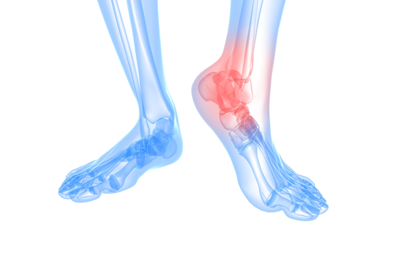
Here is a quick way to figure out if you need an X-ray after an ankle sprain:
Ankle X-ray screening questions
- Can you take four steps? (It’s okay if you limp)? No? Get an X-ray.
- Do you have tenderness/pain around your medial or lateral malleoli (the little bone bumps on either side of your ankle) – specifically the back side of these bones? Yes? Get an X-ray.
- Do you have tenderness/pain around the base of the fifth metatarsal (bump on the lateral/outside portion of your foot; halfway between your heel and your little toe)? Yes? Get an X-ray.
- Do you have tenderness/pain around the navicular bone (bump on the inside portion of your foot?) Yes? Get an X-ray.
The Ottawa ankle rules
The Ottawa ankle rules are a set of guidelines for doctors to help them decide whether a patient with foot or ankle pain should be offered X-rays to diagnose a possible bone fracture.
Before these rules, most patients with ankle injuries had x-rays taken. But the problem was, a lot of patients with unclear ankle injuries didn’t have fractures. This meant many unnecessary X-rays, which were costly, time-consuming and risky due to radiation exposure.
What happens during an X-ray
This procedure is a form of radiation like light or radio waves. It’s most commonly used after an injury to look for fractures, dislocations or bleeding in the joint.
Make sure you wear clothing that can be removed or pulled away from the joint being X-rayed.
Once an x-ray machine is carefully aimed at the part of the body being examined, it produces a small burst of radiation that passes through the body, recording an image on photographic film or a special detector.
Your doctor will place you on an X-ray table and put the x-ray film holder or digital recording plate under the table in the area of the body being imaged.
An x-ray may also be taken of an unaffected part of the body, for comparison purposes. A bone x-ray examination is usually completed within five to 10 minutes.
While a bone x-ray examination is a painless procedure, you may experience discomfort from the cool temperature in the examination room. You may also find it uncomfortable to hold still in a position and lying on the hard examination table, especially if you’re injured. Don’t worry much; your doctor will help you find a comfortable position that will help produce quality x-ray images.
After the X-ray
Depending on your condition, your doctor may advise you to go on with your daily activities or rest while you’re waiting for your results. Your results may be available on the same day as your procedure, or later.
References:
- https://theprehabguys.com/x-ray-after-an-ankle-sprain/
https://www.coreconcepts.com.sg/article/if-i-have-an-ankle-sprain-should-i-have-an-x-ray-taken/ - https://www.emedicinehealth.com/broken_ankle_or_ankle_sprain/article_em.htm#ankle_sprains_and_other_injuries
- https://www.physio-pedia.com/Ottawa_Ankle_Rules